X-ray micro-tomography
X-ray micro-tomography (micro-CT) is a non-destructive technique which is able to reconstruct the spatial 3D both outer and inner morphology of the scanned sample. The sample is irradiated by X-rays as it rotates from 0° to 360° and the 2D transmission images are taken. From these transmission images the final 3D image of the sample is then mathematically reconstructed. The advantage of this method is that no special sample preparation is required (i.e, no cutting, etching, coating, etc.). The X-ray micro-CT also distinguishes the individual phases in the material on the base of their elementary composition and of the different density of the phases. Our micro CT is Xradia MicroXCT 400 with following parameters:
- voltage of X ray source: 20 – 90 kV
- minimum voxel size: 0.2 µm
- spatial resolution < 1 µm
- maximum sample weight: 15 kg
- maximum sample diameter: 50 cm
- minimum density contrast < 10 %
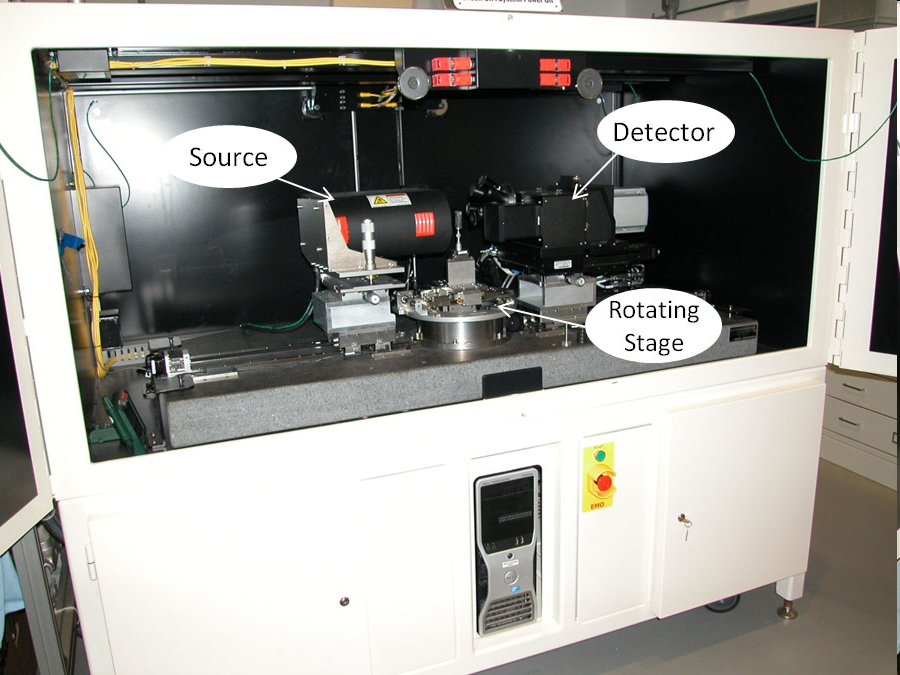
Figure 1: Xradia MicroXCT 400.
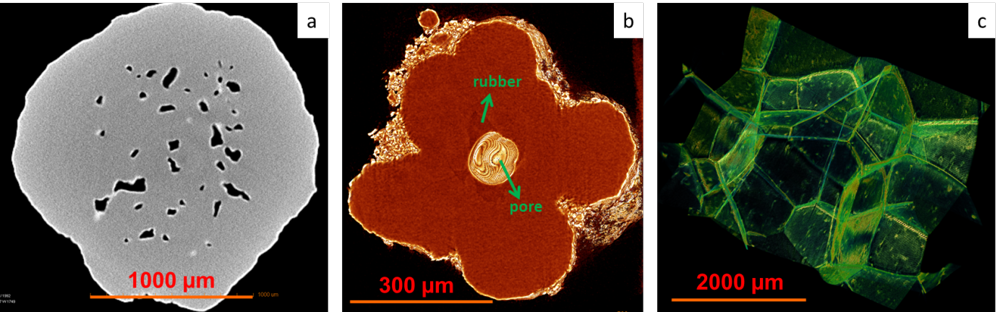
Figure 2: a) porous polypropylene particle, b) high-impact polypropylene particle with rubbery domain, c) polystyrene foam with dispersed soot.