Video-microscopy
The apparatus for the video-microcopy observation (Figure 1) consists of the observation pressure cell equipped with two glass windows and of Navitar telecentric objective with attached digital camera Nikon DS-5M. The digital camera is connected to the computer equipped with the digital image processing software LUCIA (Laboratory Imaging). The observation cell is designed as self sealing. It has the internal volume about 3 cm3 and is equipped with the capillaries for inlet and outlet of gases, temperature and pressure sensors.
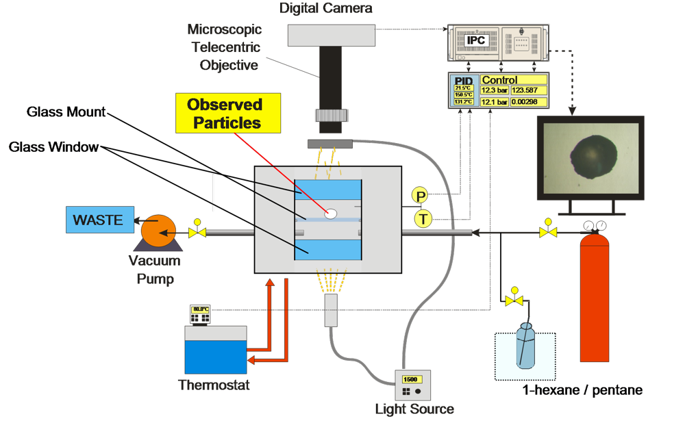
Figure 1: Schematic picture of video microscopic apparatus.
In our laboratory, the video-microscopic observation is utilized for investigation of:
- Foaming of polystyrene particles
- Polymer swelling
The polymer particle placed in the pressure cell is illuminated by the light source. The swelling/foaming polymer particle is caught by camera in the chosen interval. The change of the particle diameter in time is evaluated in the image processing software LUCIA.
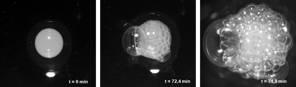
Figure 2: Foaming polystyrene particle observed in the pressure cell.